
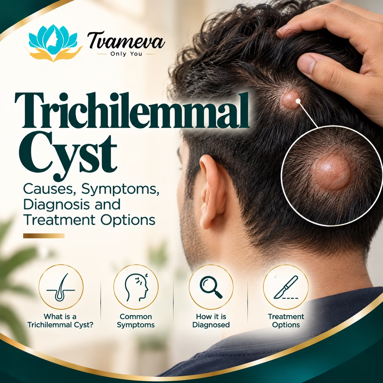
Have you noticed a firm, rounded lump beneath the skin of your scalp? While many scalp lumps are benign, they can develop for different reasons and should not automatically be assumed to be the same type of cyst.
One possible cause is a trichilemmal cyst, also known as a pilar cyst. These cysts develop from the outer root sheath of a hair follicle and are commonly found on the scalp. They may remain small and painless for years, although some gradually enlarge, become uncomfortable or cause concern because of their size or appearance.
Understanding the type of lump is important because different skin lesions can look similar but may require different approaches to diagnosis and treatment.
This guide explains what a trichilemmal cyst is, why it develops, how it differs from other cysts, when removal may be considered and what to expect from treatment. If you have noticed a persistent scalp lump, a clinical examination can help determine the appropriate next step.
What Is a Trichilemmal Cyst?
A trichilemmal cyst is a usually benign, keratin-filled cyst that develops from the outer root sheath of a hair follicle. It is also called a pilar cyst or, less commonly, a wen.
Keratin is a protein found in the skin, hair and nails. In a trichilemmal cyst, keratin accumulates within a cyst wall, creating a rounded lump beneath the skin.
These cysts most often occur on the scalp, where hair follicles are abundant. They may develop as a single lump or as multiple cysts.
A typical trichilemmal cyst is firm, smooth and mobile beneath the skin. It often has no visible opening or central punctum, which can help distinguish it from some epidermoid cysts. However, appearance alone cannot confirm the diagnosis.
Most trichilemmal cysts are benign. Rarely, a related lesion called a proliferating trichilemmal tumour can develop. A rapidly growing, ulcerated, recurrent or otherwise unusual scalp lump requires medical assessment rather than being assumed to be an ordinary cyst.
What Causes a Trichilemmal Cyst?
Trichilemmal cysts develop when keratin accumulates within a cyst formed from the outer root sheath of a hair follicle.
The exact reason an individual cyst develops is not always clear. Genetic factors can play a role, and some people have a family history of similar cysts. Multiple pilar cysts can occur in families.
1. Changes in Hair Follicle Cells
Trichilemmal cysts arise from cells associated with the hair follicle. These cells undergo a type of keratinisation that contributes to the characteristic contents of the cyst.
The resulting collection of keratin forms a rounded lump beneath the scalp.
2. Genetic Predisposition
Some families have a tendency to develop multiple pilar cysts. A family history may increase the likelihood of developing similar lesions, although not everyone with a family history will develop them.
3. Individual Skin and Follicle Characteristics
The development of a cyst is influenced by the biology of the hair follicle and the surrounding tissue. In many cases, there is no identifiable trigger.
Trichilemmal cysts should not automatically be attributed to poor hygiene, a particular hairstyle or a lack of scalp cleansing.
What Are the Symptoms of a Trichilemmal Cyst?
Symptoms depend on the cyst’s size, location and whether it has become inflamed or otherwise changed.
A Firm Lump on the Scalp
The most common feature is a rounded lump beneath the scalp. It may feel firm or smooth and may move slightly when touched.
Some cysts remain small, while others gradually increase in size.
Usually Painless
An uncomplicated trichilemmal cyst is often painless. You may first notice it while washing, brushing or styling your hair.
Pain is not a defining feature. If a lump becomes painful, tender or increasingly swollen, it should be examined.
One or Multiple Cysts
Some people develop a single cyst, while others may have several on different parts of the scalp.
Multiple lumps do not necessarily indicate a serious condition, but a clinician should assess them to confirm what they are.
Discomfort While Combing or Lying Down
A larger cyst may become uncomfortable when you brush your hair, wear headwear or lie on the affected area.
The location can also make the lump noticeable during grooming, even when it is otherwise harmless.
Redness, Swelling or Changes in the Skin
An inflamed cyst may become tender, swollen or red. Rupture can trigger an inflammatory reaction in the surrounding tissue.
Persistent redness, discharge, ulceration, rapid growth or a change in the lump’s appearance warrants medical assessment.
Where Do Trichilemmal Cysts Commonly Develop?
The scalp is the most common location for trichilemmal cysts. They can occur in different areas of the scalp and may be hidden by hair until they become larger.
A lump on the scalp is not automatically a trichilemmal cyst, however. Other possibilities include epidermoid cysts, lipomas, inflamed skin lesions and other growths.
If a lump develops outside the scalp, or if its appearance is unusual, the diagnosis should be assessed rather than inferred from location alone.
Trichilemmal Cyst vs Sebaceous Cyst: What Is the Difference?
The terms used for skin cysts can be confusing. In everyday conversation, people often use “sebaceous cyst” to describe several types of skin cysts, even though the terminology is not always medically precise.
Trichilemmal cysts and epidermoid cysts are distinct lesions, although both contain keratin and can appear as rounded lumps beneath the skin.
Trichilemmal or Pilar Cyst
A trichilemmal cyst develops from the outer root sheath of a hair follicle and most commonly occurs on the scalp. It is often firm and may not have a visible central opening.
Epidermoid Cyst
An epidermoid cyst forms from epidermal cells and contains keratin. It can develop on the face, neck, trunk and other areas. A small central opening, or punctum, may sometimes be visible.
Why the Difference Matters
The two types can look similar, and clinical examination may not always establish the exact type with certainty. If a cyst is removed, laboratory examination of the tissue may help confirm the diagnosis when indicated.
You can read more about the clinic’s existing Sebaceous Cyst Removal service to understand the assessment and treatment pathway for common skin cysts. That page addresses sebaceous or epidermoid cyst removal and is not a dedicated trichilemmal cyst service page.
Is a Trichilemmal Cyst Dangerous?
Most trichilemmal cysts are benign and do not spread to other parts of the body. A small, stable, painless cyst may not require immediate treatment if a clinician has assessed it and confirmed that observation is appropriate.
However, a lump should not be assumed to be harmless solely because it resembles a cyst.
Seek medical assessment if you notice:
- Rapid or persistent growth.
- Increasing pain or tenderness.
- Redness, warmth or swelling.
- Recurrent discharge or bleeding.
- Ulceration or changes in the skin over the lump.
- A lump that returns after removal.
- A lesion that feels fixed, irregular or otherwise unusual.
These features do not automatically mean that the lesion is malignant. They indicate that further assessment may be necessary to establish the diagnosis and determine the right treatment.
What Is a Proliferating Trichilemmal Tumour?
A proliferating trichilemmal tumour is an uncommon lesion related to the trichilemmal cyst family. It often occurs on the scalp and can present as a larger or progressively growing mass.
Many proliferating trichilemmal tumours are benign, but some can behave more aggressively. Clinical examination and histopathological assessment are important when a lesion has suspicious features.
A rapidly enlarging, ulcerated or recurrent scalp lump should not simply be monitored indefinitely as a presumed pilar cyst. A clinician may recommend removal or biopsy and laboratory examination, depending on the findings.
How Is a Trichilemmal Cyst Diagnosed?
Diagnosis begins with a medical history and physical examination. The clinician assesses the lump’s size, location, consistency, mobility, surface and rate of growth.
1. Medical History
You may be asked when the lump first appeared, whether it has grown, whether it causes pain and whether you have developed similar lumps before.
A family history of scalp cysts and any previous treatment can also be relevant.
2. Physical Examination
The clinician examines the lump and the surrounding skin. Its appearance and location may suggest a pilar cyst, but other lesions may have similar features.
3. Additional Assessment When Needed
Imaging is not necessary for every typical superficial cyst. However, additional assessment may be recommended if the lump is deep, unusually large, fixed, rapidly growing or difficult to diagnose clinically.
4. Histopathological Examination
If a cyst or suspicious lesion is removed, the tissue may be sent to a laboratory for histopathological examination. This allows a pathologist to assess the tissue under a microscope and confirm the diagnosis when indicated.
The need for laboratory examination depends on the clinical findings and the treating clinician’s judgement.
Does a Trichilemmal Cyst Need Treatment?
Not every trichilemmal cyst needs to be removed. The decision depends on the diagnosis, symptoms, growth pattern and individual preferences.
When Observation May Be Appropriate
A small, stable and painless cyst may be monitored if a clinician has assessed it and is comfortable with observation.
Monitoring should not mean ignoring changes. If the lump grows, becomes painful or develops new features, it should be reassessed.
When Removal May Be Considered
Removal may be considered when a cyst:
- Continues to grow.
- Causes discomfort or interferes with grooming.
- Becomes repeatedly inflamed.
- Creates a cosmetic concern.
- Is repeatedly irritated by combing or headwear.
- Has an uncertain diagnosis.
- Has features that warrant tissue examination.
The choice should be made after an assessment of the lump and a discussion of the expected benefits, risks and possible scar.
How Is Trichilemmal Cyst Removal Performed?
When removal is appropriate, surgical excision is commonly used. The exact technique depends on the cyst’s size, location, condition and relationship to the surrounding tissue.
Step 1: Examination and Treatment Planning
The clinician assesses the cyst and determines whether removal is suitable. If the lesion is inflamed, infected or unusually large, the treatment plan may need to be adjusted.
You should be informed about the proposed procedure, expected healing, possible complications and whether the removed tissue will be sent for examination.
Step 2: Anaesthesia
Many small, superficial cysts can be removed using local anaesthesia. The approach may differ for larger, deeper or more complex lesions.
Local anaesthesia numbs the area so that the procedure can be performed with reduced discomfort.
Step 3: Surgical Removal
The surgeon makes an incision appropriate to the size and location of the cyst and removes the lesion. When feasible, removing the cyst wall helps reduce the chance of recurrence.
The incision may be closed with stitches or managed using another suitable wound-closure method.
Step 4: Tissue Examination When Indicated
If histopathological examination is recommended, the removed tissue is sent to a laboratory. The clinician will explain how and when results will be communicated.
Step 5: Wound Care and Follow-Up
The treated area is dressed as required, and you receive instructions on keeping it clean, protecting the wound and attending follow-up.
If stitches are used, a follow-up appointment may be required to assess healing and remove non-dissolvable sutures when appropriate.
Can a Trichilemmal Cyst Be Removed Without Surgery?
Treatment depends on the diagnosis and the cyst’s characteristics. There is no reliable home remedy or topical cream that can be expected to remove the entire cyst wall.
Squeezing, puncturing or attempting to drain a cyst at home may cause irritation, infection, bleeding or rupture. It can also make later assessment and treatment more difficult.
If removal is appropriate, a clinician can explain the available procedural options. The choice should prioritise correct diagnosis and safe removal rather than attempting to eliminate the lump with unproven remedies.
Can a Trichilemmal Cyst Come Back After Removal?
Yes, recurrence is possible, particularly if part of the cyst wall remains. Removing the cyst and its wall when feasible reduces the chance of recurrence, but it cannot guarantee that a cyst will never return.
A new lump near the previous site may represent recurrent cyst tissue, a separate cyst or another type of skin lesion. It should be examined rather than assumed to have the same cause.
If a lump returns repeatedly, changes rapidly or looks different from the original lesion, further evaluation may be needed.
What Is the Recovery Time After Trichilemmal Cyst Removal?
Recovery depends on the cyst’s size, the incision, the closure method and individual healing.
After a straightforward procedure, you may experience mild tenderness, swelling or bruising around the treated area. These symptoms should generally improve rather than progressively worsen.
Your clinician will explain how to care for the wound, when you can wash your hair, whether you need to avoid strenuous activity and when to return for review.
If non-dissolvable stitches are used, they may need to be removed at an appropriate follow-up appointment. Do not remove them yourself.
The wound may close before the scar has fully matured. Scar colour and texture can continue to change for several months, and the final appearance varies between individuals.
For more information about wound care and recovery after cyst removal, read the existing guide to Sebaceous Cyst Removal Recovery. Its advice relates to general cyst-removal recovery; your own surgeon’s instructions should take priority.
What Are the Risks of Trichilemmal Cyst Removal?
As with any minor surgical procedure, cyst removal has potential risks. These vary according to the cyst’s size, location, condition and the procedure performed.
Possible risks include:
- Bleeding or bruising.
- Infection.
- Temporary tenderness or swelling.
- Delayed wound healing.
- A visible scar.
- Incomplete removal or recurrence.
- A need for additional assessment or treatment if the diagnosis is uncertain.
A cyst that is inflamed or infected may require a different approach from an uncomplicated cyst. Your clinician should explain the risks that are most relevant to your situation.
Seek medical advice if you develop increasing redness, warmth, swelling, worsening pain, pus, fever, persistent bleeding or wound separation after treatment.
How Much Does Trichilemmal Cyst Removal Cost in Mumbai?
The cost depends on several factors, including:
- The cyst’s size and location.
- Whether there is inflammation or infection.
- The complexity of removal.
- The type of anaesthesia and facility required.
- Wound closure and follow-up needs.
- Whether histopathological examination is recommended.
A reliable estimate requires an examination of the lump and confirmation of the proposed treatment. Ask what the quoted fee includes and whether laboratory testing or follow-up appointments are charged separately.
Do not choose a treatment based on cost alone. Correct diagnosis, appropriate surgical technique and safe follow-up are important considerations.
When Should You Consult a Doctor About a Scalp Lump?
Arrange a clinical assessment if you notice a new or persistent lump on your scalp, especially if it is growing or changing.
Seek prompt medical advice if the lump becomes increasingly painful, red, warm or swollen, or begins discharging fluid. A rapidly enlarging, bleeding or ulcerated lesion also requires timely evaluation.
Avoid squeezing or cutting the lump yourself. An examination can help establish whether it is likely to be a pilar cyst or whether another diagnosis needs to be considered.
Trichilemmal Cyst Assessment at Tvameva Aesthetics, Vile Parle West
If you have noticed a persistent scalp lump, a clinical assessment can help determine its likely cause and whether observation, further investigation or removal may be appropriate.
Dr. Kunal Sayani, MBBS, MS, MCh and DrNB (Plastic Surgery), provides surgical consultations at Tvameva Aesthetics in Vile Parle West, Mumbai. The appropriate treatment depends on the examination and the nature of the lesion.
You can read about the clinic’s existing Sebaceous Cyst Removal service for information about its general cyst-removal pathway. Please note that this is not a dedicated trichilemmal cyst service page; confirm the availability of assessment and treatment for your specific concern with the clinic.
To enquire about a consultation, visit the Contact page.
Frequently Asked Questions About Trichilemmal Cysts
1. What is another name for a trichilemmal cyst?
A trichilemmal cyst is also known as a pilar cyst. It develops from the outer root sheath of a hair follicle and is most commonly found on the scalp.
2. Are trichilemmal cysts cancerous?
Most trichilemmal cysts are benign. Rarely, a related proliferating trichilemmal tumour can behave more aggressively. A growing, ulcerated, recurrent or unusual lesion should be examined and investigated when indicated.
3. Can a trichilemmal cyst go away on its own?
A cyst may remain stable for years, but it generally does not disappear simply because it is left alone. If it is small and has been assessed as benign, observation may be appropriate. Treatment decisions should be based on the diagnosis and symptoms.
4. Are pilar cysts painful?
They are often painless. Pain or tenderness can occur if a cyst becomes inflamed, ruptures or is irritated. Increasing pain or swelling should be assessed by a clinician.
5. How is a pilar cyst different from an epidermoid cyst?
A pilar cyst develops from the outer root sheath of a hair follicle and commonly occurs on the scalp. An epidermoid cyst develops from epidermal cells and can occur in several areas of the body. Both contain keratin and can look similar, so examination may be needed to distinguish them.
6. Is surgery necessary for every trichilemmal cyst?
No. A small, stable, painless cyst may be monitored after appropriate assessment. Removal may be considered if it grows, causes symptoms, affects appearance or has features that require further investigation.
7. Can a pilar cyst return after removal?
Yes. Recurrence can occur if part of the cyst wall remains. Complete removal of the cyst wall when feasible reduces the likelihood of recurrence but does not eliminate the possibility entirely.
8. Does removing a trichilemmal cyst leave a scar?
Any procedure involving an incision can leave a scar. Its appearance depends on the size and location of the cyst, the surgical technique and individual healing. No technique can guarantee an invisible scar.
9. Can I squeeze or drain a trichilemmal cyst at home?
No. Squeezing or puncturing a cyst may cause inflammation, infection, bleeding or rupture and may make later treatment more difficult. Seek medical assessment instead.
10. When should I have a scalp lump examined?
Arrange an assessment for a persistent or growing lump. Seek prompt advice if it becomes painful, red, swollen, ulcerated or starts bleeding or discharging fluid. Rapid growth or repeated recurrence should not be ignored.
Final Thoughts
A trichilemmal cyst, or pilar cyst, is usually a benign growth that commonly develops on the scalp. Although many remain painless and stable, the appearance of a lump alone cannot confirm its diagnosis.
A clinical assessment can help distinguish a pilar cyst from other scalp lesions and establish whether observation, further investigation or surgical removal is appropriate. When removal is recommended, the approach should consider the lesion’s characteristics, the possibility of recurrence, wound healing and whether tissue examination is needed.
If you have a persistent or changing scalp lump, seek an assessment rather than attempting to drain it at home. An individualised plan can help you understand the available options and make an informed decision about treatment.
Leave a Reply